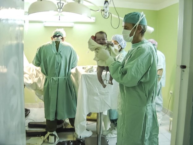
Right Shunt Congenital Disease Early Cyanosis Blue Babies Usmle

Definition and Background
Cyanosis refers to the condition where the tissues impart a bluish or purplish discoloration due to reduced tissue perfusion or reduced blood oxygenation.
For the cyanosis to be appreciated clinically in a neonate, approximately 3 g/dL of deoxygenated hemoglobin should be present in the capillaries to generate the dark blue color.
Cyanosis is a very frequent outcome in newborn babies. Neonatal cyanosis, especially of the central type, can result due to significant and possibly life-threatening conditions related to the cardiopulmonary, metabolic, neurological conditions, as well as infections.
Types of Cyanosis
Central cyanosis
Central cyanosis occurs due to decreased oxygenation of hemoglobin.
It is common in newborn babies and resolves within the first 10 minutes after birth as lungs expand and cardiopulmonary physiology changes after birth. However, persistent central cyanosis is usually abnormal and is due to cardiac or respiratory issues that prevent proper oxygenation. It must be assessed and treated immediately.
Peripheral cyanosis
In peripheral cyanosis, there is a normal systemic arterial oxygen saturation. But cyanosis still occurs in the peripheral tissues due to increased uptake of oxygen by the tissues resulting in an enlarged systemic arteriovenous (AV) oxygen difference and increased deoxygenated hemoglobin concentration in the venous blood.
This increased oxygen extraction by the tissues and a wide systemic AV oxygen difference may occur due to a variety of factors that slow down the blood flow through the capillaries, for instance:
- Elevated venous pressure
- Low cardiac output
- Polycythemia
- Vasoconstriction (caused by cold exposure, drugs, connective tissue disorders)
- Vasomotor instability
- Venous obstruction
Peripheral cyanosis is characteristically seen in the distal part of the extremities and less frequently around the circumoral or periorbital regions.
Acrocyanosis
Acrocyanosis isbenign peripheral cyanosis surrounding the mouth and extremities (i.e. hands and feet) in healthy newborns. It occurs due to benign vasomotor alteration resulting in peripheral vasoconstriction. It is usually a common presentation and may likely to last up to 48 hours.
Causes of Cyanosis
Central Cyanosis
In neonates, central cyanosis results due to significant respiratory, cardiac, or circulatory disorders that prevent tissue oxygenation. The important causes are highlighted below:
Cardiac causes:
- Transposition of great arteries
- Tetralogy of Fallot
- Total anomalous pulmonary venous return
- A left heart that is small or hypoplastic
- Truncus arteriosus
- Persistent fetal circulation
Respiratory causes:
- Birth trauma or asphyxia
- Transient tachypnoea of the newborn
- Respiratory distress syndrome
- Pneumothorax
- Pulmonary or lung edema
- Accidental aspiration or swallowing and choking on meconium
- A diaphragmatic hernia
- Pleural effusion
- Trachea-oesophageal fistula
- Obstruction of the upper respiratory tract
Other causes:
- Low blood sugar
- Inadequate blood magnesium
- Infections
- Epilepsy
Peripheral cyanosis
The causes of peripheral cyanosis include:
- Central cyanosis and all its causes
- Decreased cardiac output
- Defects in the circulation of the blood, such as thrombosis or embolism
- Reduction in the size of blood vessels of the arms, legs, fingers, and toes due to:
- Exposure to cold
- Raynaud's phenomenon
- Spasm of the tiny skin capillaries or arteries referred to as acrocyanosis
- Erythrocyanosis taking place in young women or as an unwanted effect of beta blocker drugs taken for hypertension
Clinical Features
As the name suggests, the patients with cyanosis have a bluish discoloration of the skin and mucous membranes, commonly affecting the lips, extremities, fingers, and toes. The other symptoms depend upon the underlying disease that has precipitated the cyanosis. For example:
- Tachycardia, tachypnea, fever, cold extremities, in cases of septic shock
- Cardiac murmurs, edema, in cases of congenital heart diseases
- Crepitations, wheezes, grunting sounds, dyspnea, tachypnea, shallow breathing in cases of respiratory diseases
Additionally, neonates born with cyanosis are of low weight. They are likely to get tired easily and sweat profusely while eating. They may probably be irritable and sometimes feel floppy or weak. There is a flaring of the nose revealing that the baby is breathing with stress (labored breathing). They also develop frequent infections.
Diagnosis
Although cyanosis is a clinical diagnosis, a proper history and examination are important to shortlist the etiology of cyanosis.
History
A detailed history of prenatal care, pregnancy, labor, birth trauma, and infant risk factors should be obtained. If the mother has a history of gestational diabetes, infections or smoking, this heightens the risk of developing congenital heart disease.
Presence of oligohydramnios may point to renal abnormalities related to hypoplastic lungs, while polyhydramnios is likely to show airway, esophageal, or neurological abnormalities.
A history of prolonged labor may have led to intracranial hemorrhage or phrenic nerve paralysis.
Clinical Examination
In physical examination, the pattern and features of growth should be noted because babies who are either too small or too large for their age are more likely to develop polycythemia. The main aim is to determine the extent of respiratory distress because it will point to congenital heart disease or methemoglobinemia.
A lack of adequate respiration caused by the pulmonary disease is naturally revealed by fast respiration associated with chest retractions and nasal flaring.
Neurological diseases may lead to cyanosis owing to hypoventilation and may cause reduced or irregular respirations. It is also imperative that the baby's breathing tone and activity are evaluated to check for periodic breathing and/or the presence of apnea.
The cardiac examination must include an evaluation of the baby's heart rate, peripheral pulses, as well as perfusion. While auscultating the heart, concentrate on the second heart sound, which is likely to be loud and heard once in pulmonary hypertension. Carrying out auscultation for cardiac murmurs is important.
Investigations
Laboratory investigations
A complete blood count to look for polycythemia and infections.
Pulse oximetry should be carried out to determine the level of oxygen saturation. It is important that measurement should be obtained from both the hand and a foot to ascertain the pattern of flow via the ductus arteriosus.
While a venous blood gas is important for evaluating pH and PaCO2, it should not be utilized to ascertain oxygenation. However, if significant metabolic acidosis is revealed, it may point to cardiac failure, sepsis, asphyxia, or metabolic disorders.
Chest Radiograph
A chest radiograph is a very vital part of evaluating a cyanotic baby. It helps to look at the lung fields as well as positions of stomach, liver, and heart, It will also help to eliminate dextrocardia and situs inversus.
Elevation of a hemidiaphragm by more than two intercostal spaces in comparison to the other side points to diaphragmatic paralysis caused by phrenic nerve injury.
Reduced pulmonary vascular outlines suggest pulmonary stenosis or pulmonary atresia.
The size and shape of the heart are likely to help point to a diagnosis: the heart is "boot-shaped" in the tetralogy of Fallot, while "egg on a string" sign is seen in the transposition of great arteries.
Electrocardiogram (ECG)
An electrocardiogram (ECG) is important to diagnose cardiac arrhythmias, abnormal cardiac axis, and ventricular hypertrophy. Though ECG is not very useful in assessing a baby with congenital heart disease because it may appear totally normal even in life-threatening cardiac conditions, such as transposition of great arteries.
Hyperoxia Test
The hyperoxia test is an important clinical tool for differentiating pulmonary conditions from cardiac diseases in cyanotic babies.
It is based on the theory that if there is an absence of persistent cardiac shunts, then administration of 100 % oxygen will increase blood PaO2, while in babies with cyanotic congenital heart disease, we will see no observable difference in the amount of PaO2 even after breathing 100% O2.
- 100 % FiO2 for 10 minutes
- Draw an ABG
- > 200 mm Hg: cardiac disease unlikely
- 50—150: suggest mixing lesion (truncus, tricuspid atresia)
- < 50: suggests two circuits with mixing (TGA)
Source: https://www.lecturio.com/magazine/blue-baby-syndrome/
0 Response to "Right Shunt Congenital Disease Early Cyanosis Blue Babies Usmle"
Postar um comentário